How to do Lung Ultrasound to rule out Pneumothorax!
1. Select your probe:
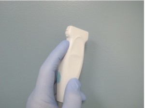
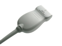
Linear probe, or vascular probe with low penetration and high frequency.
2. Technique:
Transducer should be in longitudinal position. It means your orientation marker should be toward the head of patient. Patient should be in supine position and probe should be in inter-coastal areas and you should scan at least 3 inter-coastal spaces. You can start from ant. axillary line toward lateral sternal border, or mid clavicle line, with this position you are looking at the pleura.
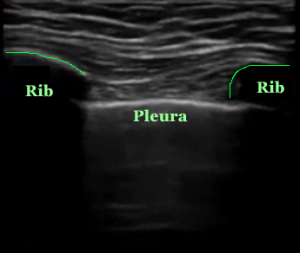
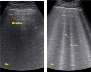
Pleura is a bright whit line between two ribs. and if pleura moves with respiration , you should see a vertical line below the pleura. This line is an artifact (comet tail) and represents sliding of pleura(Fig.1). This artifact should be differentiate from B-Line. B line is a vertical lines that arises from the pleural line and continues to the edge of the screen.(Fig.2) B line will rule out pneumothorax. If we do not see any lung sliding and comet tail artifact, we should be suspicious for pneumothorax.
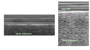
If you do not see comet tail(reverberation) artifact and you would like to to have a confirmation test for that, you can proceed with M-Mode. you will set your M-Mode line in the middle of 2 ribs to go through pleural line. If you have a normal lung, M- Mode should show you some wavy, grainy, “sandy beach” appearance (seashore sign).
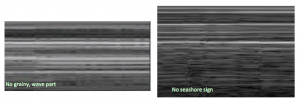
If M-Mode show you no seashore sign, means there is no motion on pleura and there is no pleural sliding so you will see the following images = PTX
I do not want to confuse you but there are other lines in lung ultrasound such as A line, Z line , and E line. Just for your info, A lines are horizontal lines parallel to pleural line, with same bright echogenic feature.If there are A lines in the absence of lung sliding this can represent Pneumothorax. If A lines presents with lung sliding this could be Asthma or COPD.
